
Amos's operation on 11 June 2026 took eight hours and twenty minutes, by Pride's own note. The post-operative radiographs started at 20:22.
DICOM (Digital Imaging and Communications in Medicine) is the international standard for storing, transmitting, printing, and managing medical imaging information and related data
A DICOM file doesn't just contain an image, its a log: every exposure carries the time it was taken, the protocol selected, the exposure time in milliseconds, and the detector's exposure index, which is the machine's own measure of whether enough X-rays reached the plate.
Pride sent me 6 DICOM files for Amos' post-op radiographs.
| Time | View | Exposure | Exposure index |
|---|---|---|---|
| 20:22 | Lateral | 4 ms | 83 |
| 20:35 | VD | 5 ms | 142 |
| 20:36 | VD | 5 ms | 134 |
| 20:45 | VD | 5 ms | 123 |
| 20:54 | Lateral | 27 ms | 173 |
| 20:57 | VD | 35 ms | 158 |
A lateral is taken from the side, with the dog lying on his flank. A VD, ventrodorsal, is taken with the dog on his back: the beam enters through the belly (ventral) and leaves through the spine (dorsal) onto the plate beneath him. Amos’s construct sits at the lumbosacral junction, where his lower back joins his pelvis, a hand’s width in front of where his tail starts.
The DICOM headers carry instance numbers. In the post-op series: the numbers run from 1001 to 1009, so three exposures were taken and deleted before export.
The protocol on every film is LARGE_DOG_PELVIS. The films taken on 3 July, on the same machine, use LARGE_DOG_LUMBAR, which is the protocol for a lumbosacral construct, which is what Amos has. The operator field on all six reads USER.
20:22
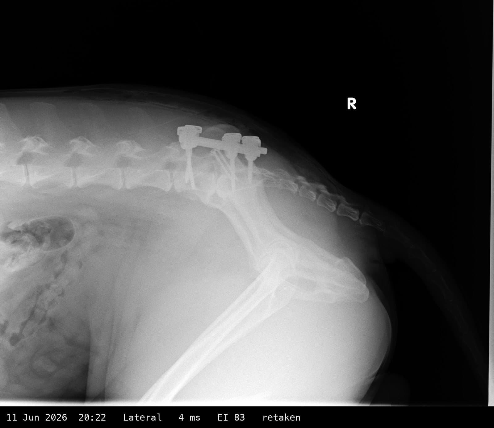
The first film of the night. Four milliseconds is a tiny exposure for of Amos' size: the detector's own index of 83 says almost nothing reached it. On the picture that shows as a washed-out spine and a construct you can make out only because titanium is bright at any dose.
Look at the shape of him. His back legs are pulled forward under his belly, thigh bone nearly at right angles to the spine. The spine itself arches up over something he's lying on, shown by his tail falling downwards and away from his body. The two sides of the pelvis don't sit on top of each other, so you can see a double outline where there should be one. For a side view of a joint that was fixed straight not even an hour earlier, every one of those is the opposite of what you want: the joint is bent, the pelvis is twisted, and the picture is too faint to read.
Thirteen minutes pass before the next exposure.
20:35
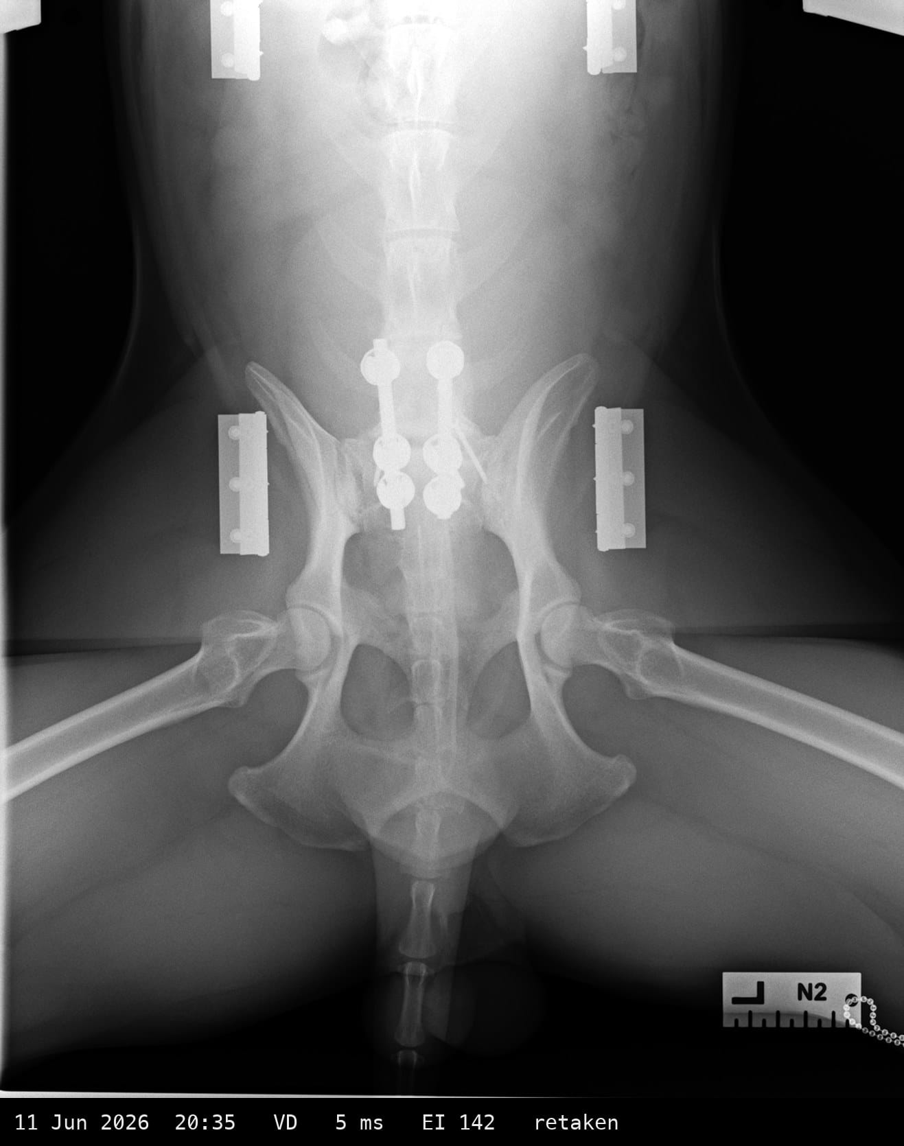
Belly-up now, which means Amos is lying on the wound that was just closed, with the metalwork between his spine and the table. The exposure has gone up by a single millisecond. The index says still underexposed.
Three things in the picture that aren't Amos: four rectangular metal brackets at the corners, two beside the hips and two cut off at the top, which are the fittings of whatever he's lying in; a hard horizontal line across the film at hip level, where a pad under his pelvis ends; and the letter N2 on a ruler in the corner, the marker.
Now look at where the metalwork is: the middle third of the picture. Everything below it is his pelvis and his back legs, which have simply been left where they fell when he was rolled onto his back, splayed out to either side. Nobody has positioned him.
20:36
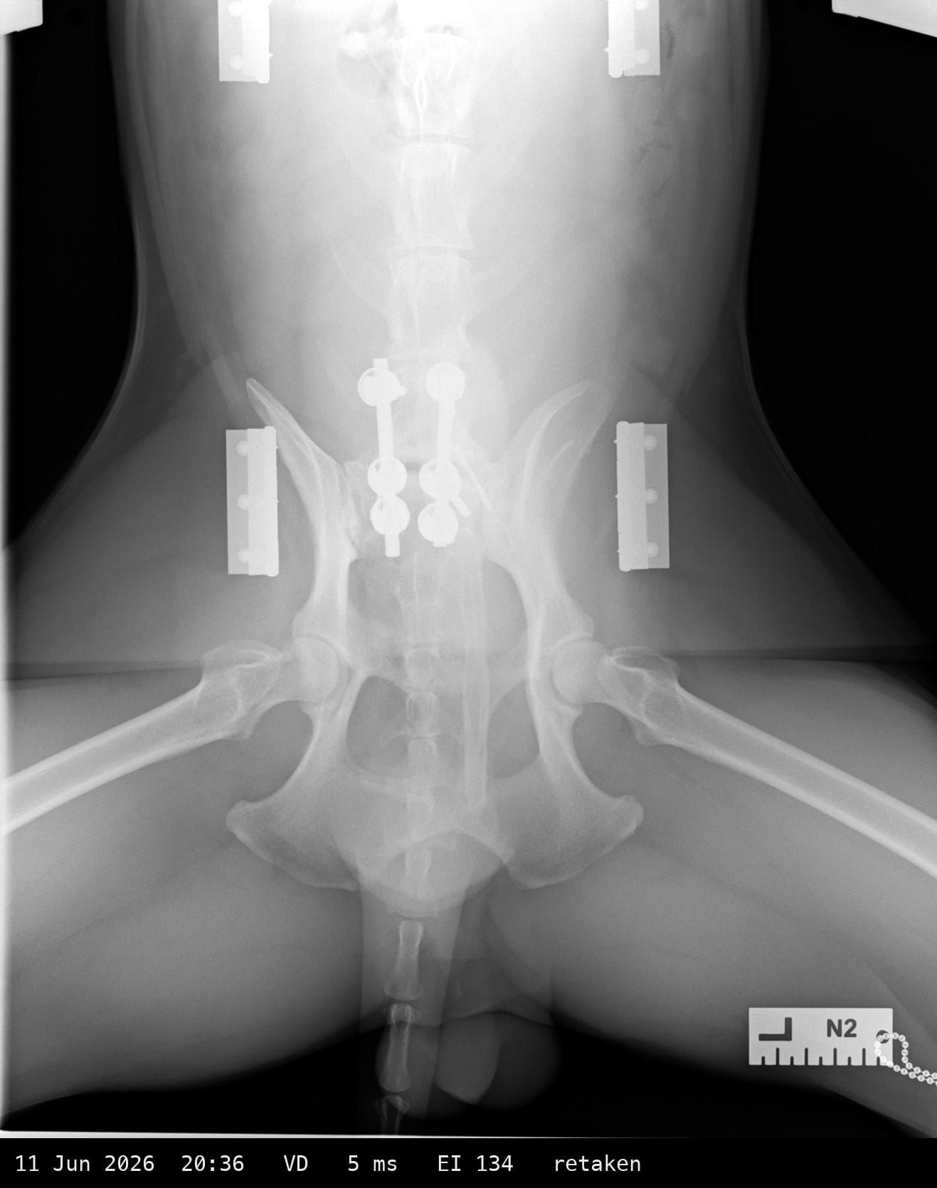
One minute later. Same exposure, same position, same brackets, same pad line. Nothing has been changed except that the picture was taken again. The index came out slightly lower.
If at first you don't succeed, try exactly the same thing again?
20:45
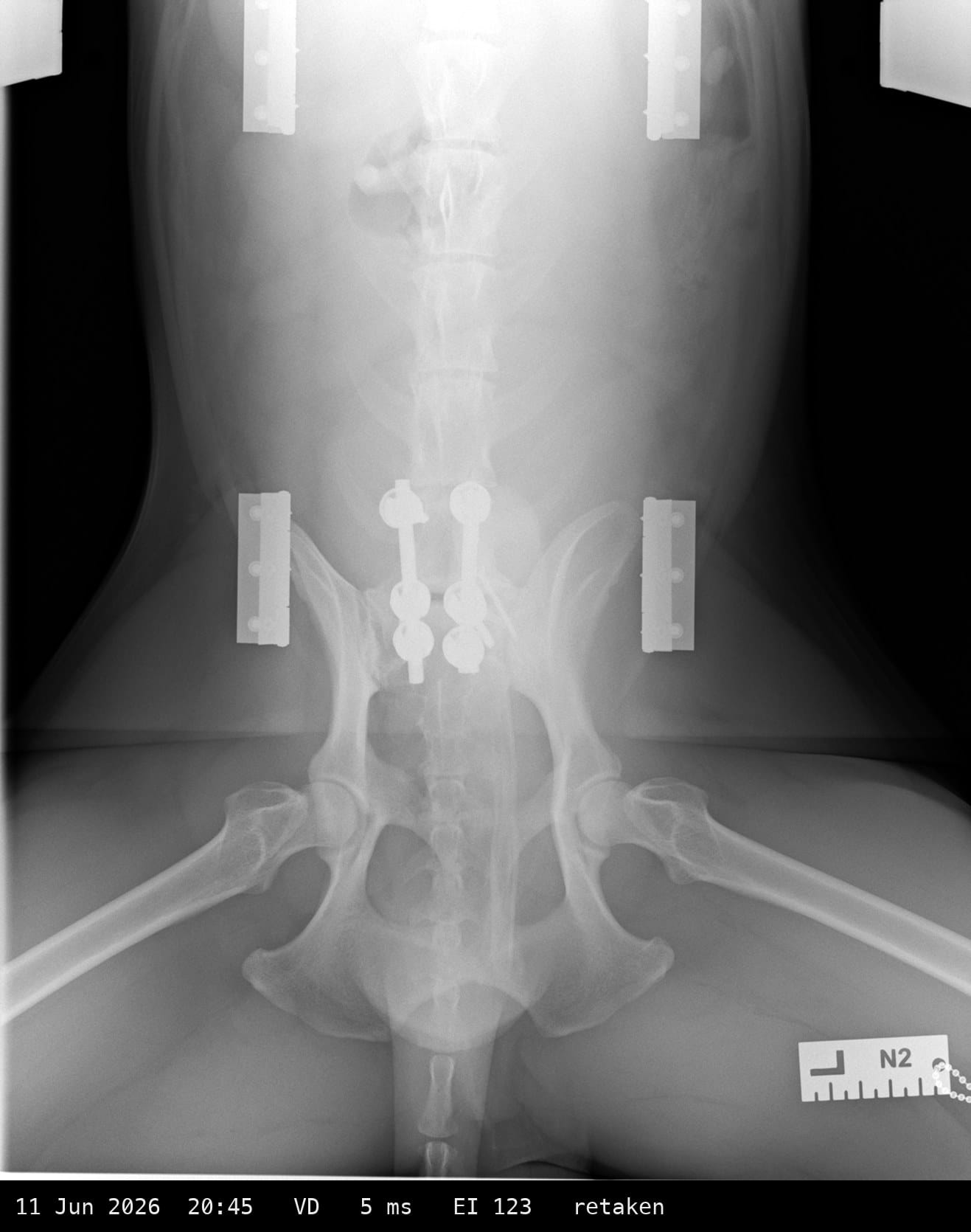
Nine minutes later. Still 5 ms. The framing has moved: the brackets at the top are now fully in view, so the beam or the dog has shifted down the table, but the dog hasn't been repositioned and the dose hasn't been touched. This is the third attempt at the same setting and the index is the lowest of the three.
This would almost be comical, if I weren't writing about my own dog.
20:54
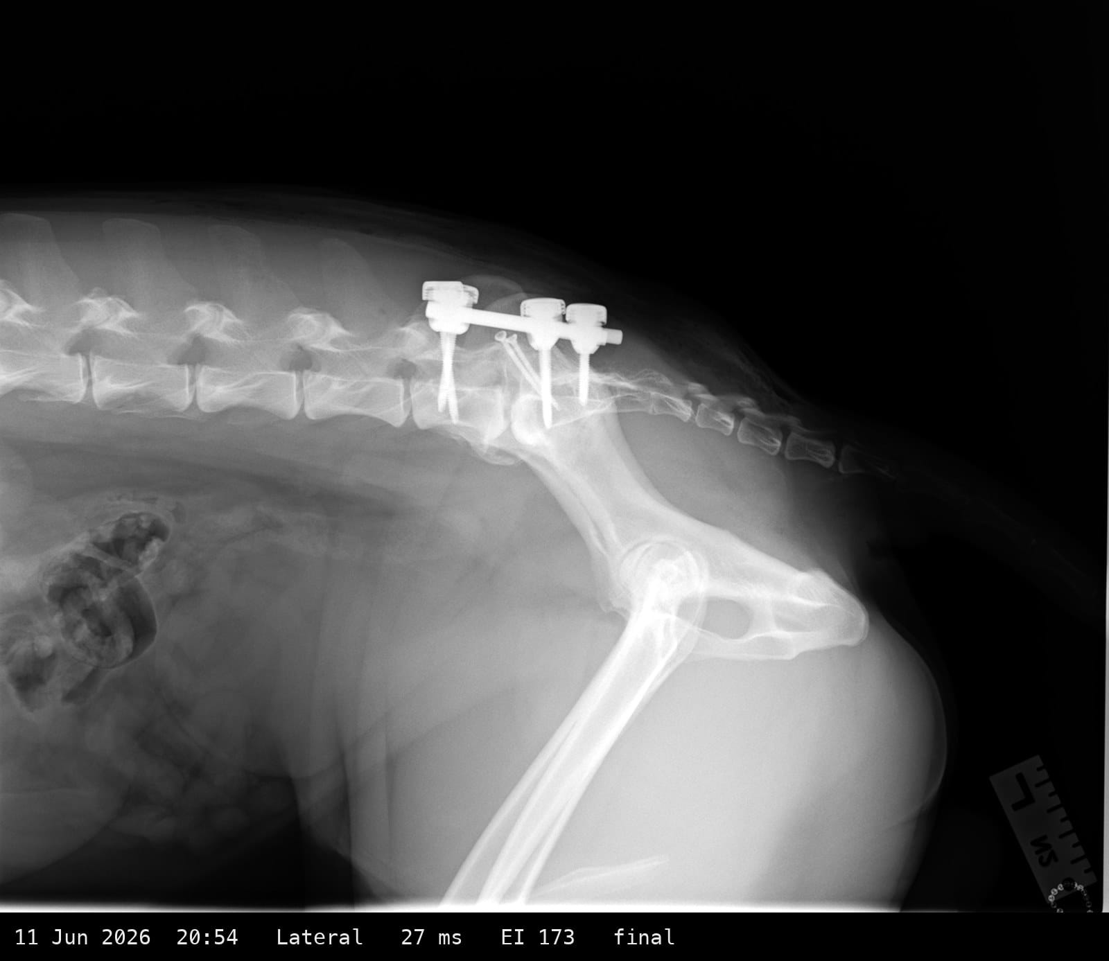
Nine minutes after the last failed VD, the exposure goes from 4 ms to 27 ms, nearly seven times the first attempt, and the lateral comes out readable first time.
I think someone who knew what they were doing entered the room, or was finally asked. Thirty-two minutes late.
20:57
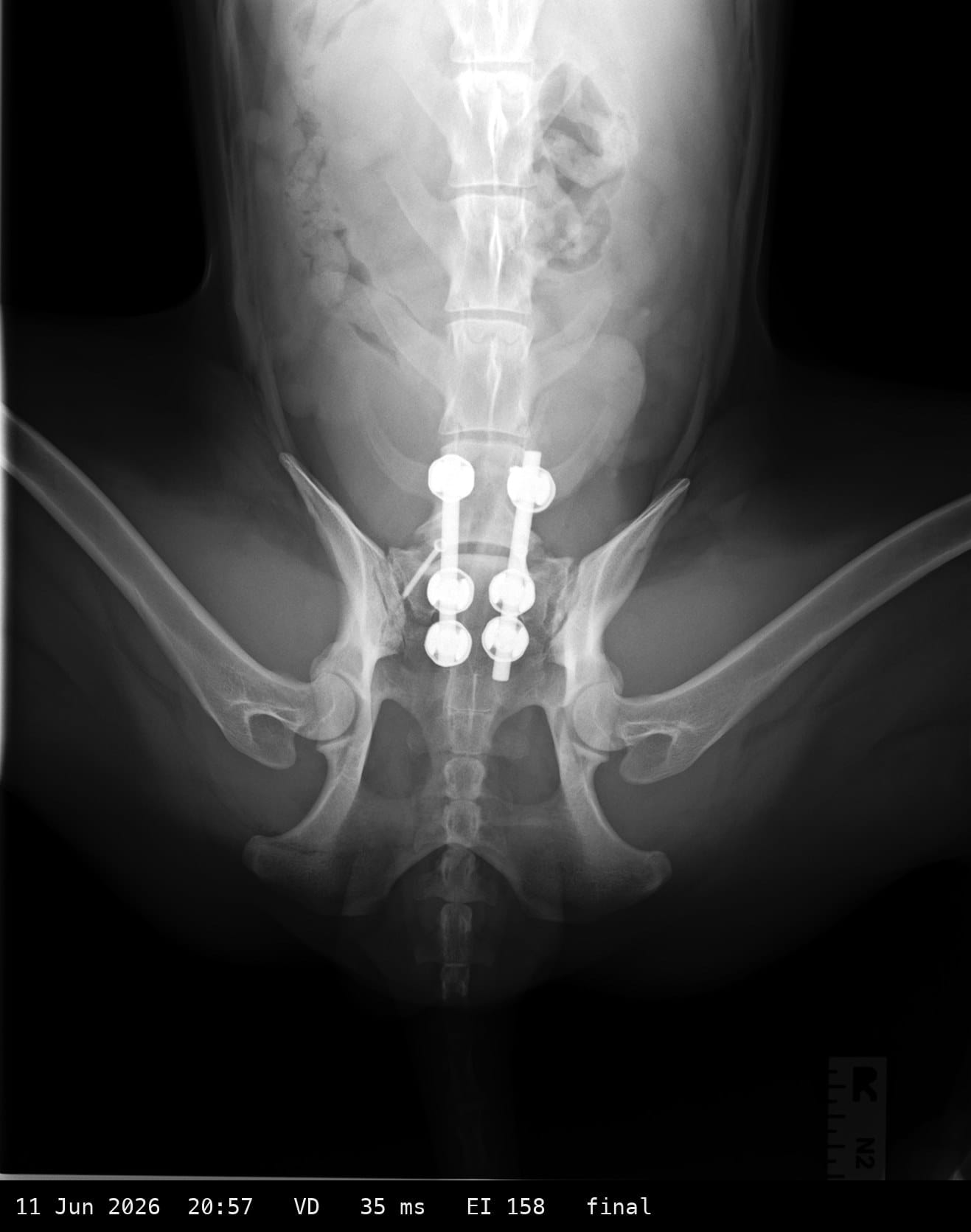
Three minutes later, the last film. This one is different in every way that matters. The brackets are gone. The pad line has moved. The metalwork is in the centre of the picture. The pelvis sits square, both sides matching. His legs are out to the sides instead of hanging naturally. Somebody lifted him out of the cradle, lined the beam up on the joint instead of the hips, and set the exposure.
Done properly, the two views took three minutes.
He had been on the x-ray table for thirty-five, after an eight hour twenty minute surgery. And that’s only counting from the first exposure.
Nine exposures over 35 minutes to produce two usable pictures. Four attempts at the wrong dose with the dog never moved between them. A single intervention at 20:54 that produced two usable images in three minutes.
Since July I have asked Pride, and then IVC Evidensia, for the records of that evening: the anaesthetic record, the theatre chart, who was in the room. It is 20 October. What I have is six files with “USER” in the operator field, and the bill.